
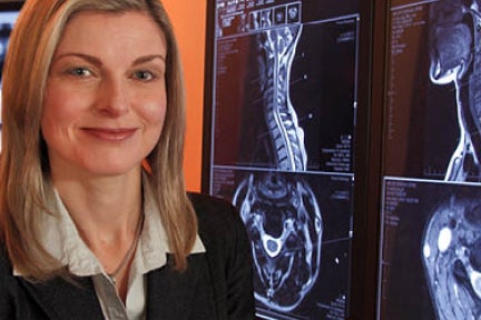
Magnetic Resonance Imaging (MRI)
Superlative soft tissue detail makes MR the preferred modality for diseases of the brain including stroke, inflammatory conditions such as multiple sclerosis, and degenerative conditions. MR allows accurate assessment of the intervertebral discs, spinal and peripheral nerves, and the bone marrow and soft tissues in and around joints.
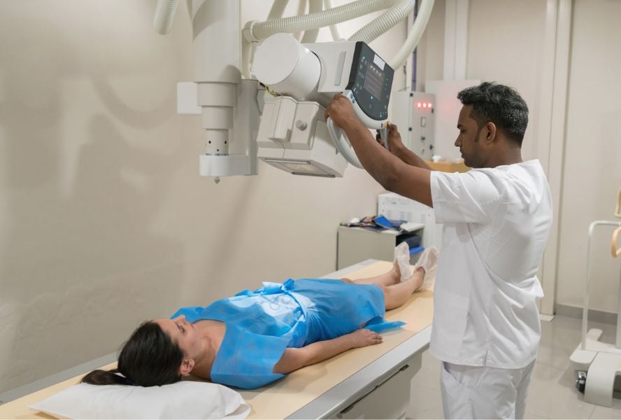
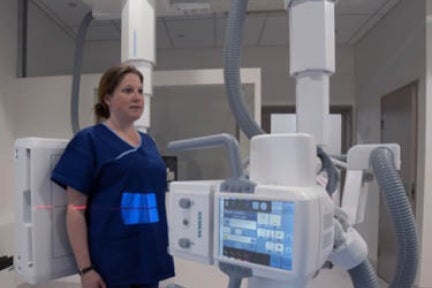
X-ray
X-rays are a quick and efficient modality for assessing fractures, the lungs, cardiac contour, and seeing soft tissue calcification. Our clinics offer state-of-the-art digital general x-ray imaging. Our expert technicians also use x-ray as an accurate method for assessing breast cancer (mammography), and in barium studies of the gastrointestinal tract.

Ultrasound
Ultrasound uses high-frequency sound waves to give anatomical and functional imaging of soft tissues. It is safe, fast and capable of very high resolution. Examples of ultrasound use include gynaecology and obstetrics, abdomen viscera, breast, pelvis, thyroid, testes, and the musculoskeletal system. Doppler ultrasound is used to asses flow and patency of arteries. Ultrasound is used to guide interventional procedures.

Diagnostic mammography
Our low-dose mammograms are used in assessing and screening breast pathologies. The equipment is the latest digital technology, providing the best possible pictures.
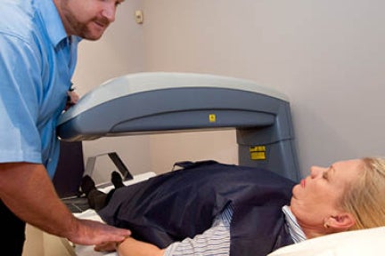
Bone mineral density
Bone densitometry or dual-energy x-ray absorptiometry (DEXA) is used to assess and monitor the presence of osteoporosis. Practitioners rely on our scans to provide information about bone strength or fragility, to help in monitoring bone loss and in planning any preventative therapy or medical treatment.
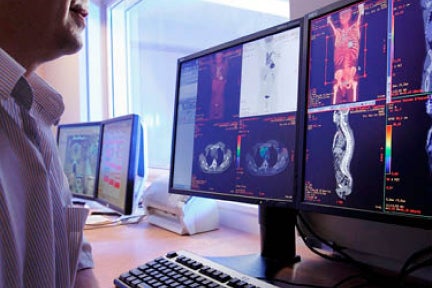
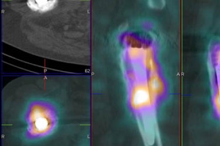
Positron Emission Tomography and Computed Tomography (PET/CT)
PET/CT uses radiopharmaceuticals to diagnose, stage and monitor a variety of malignancies, and to assess the metabolic activity of a variety of disease processes. It is also useful in assessing cardiac function and neurodegenerative disease. The “metabolic” image is fused with a CT image to allow high anatomical detail.
Nuclear medicine
Nuclear medicine tests use radiopharmaceuticals to assess metabolic function - especially in the skeleton. They are also used to assess heart disease, gastrointestinal, endocrine and neurological disorders.
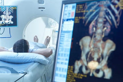
Computed Tomography
The new generation of CT scanners are fast and precise, delivering anatomical and functional detail at a relatively low radiation dose. This is the optimal modality for assessing the spine, lungs, mediastinum, abdomen and pelvis. Because of its speed, CT is ideal for assessment of complications following trauma, including brain haemorrhage. CT angiography is a non-invasive method to assess vessels.
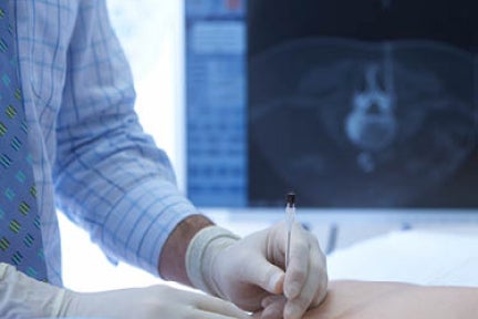
Interventional procedures
Interventional radiology utilises a variety of image-guided techniques (mostly CT and ultrasound). Our interventional radiologists perform biopsies, treatment for varicose veins, and therapeutic and diagnostic injections into joints, tendons and around inflamed nerves.
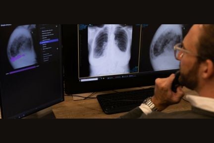
Artificial intelligence solutions
Annalise.ai is a joint venture between healthcare technology company Harrison.ai, and I–MED Radiology. This partnership amplifies the potential of clinical diagnosis through AI, aiming for enhanced health outcomes. Within I-MED Radiology, cutting-edge AI solutions, such as Annalise CXR and Annalise CTB, empower radiologists in identifying chest and neurological diseases. This innovation holds promise for improving patient health outcomes significantly.
New and evolving imaging systems
I-MED Radiology Network is at the forefront of new and evolving imaging systems and methods. Some examples are the standing MRI units, dedicated erect feet imaging and full length body imaging.