Ultrasound
Ultrasound
What is an ultrasound?
An ultrasound examination is performed using a smooth, hand held device called a transducer (camera) that moves across the body with a sliding and rotating action. The transducer transmits high-frequency sound waves into your body. The sound waves are then reflected from the different tissues in different ways and converted to electrical impulses, which are used to produce a moving image onto the screen.
While ultrasound is a great diagnostic tool for soft tissue, superficial imaging, it cannot be used for all areas of the body - some structures/areas may be better seen with X-rays, CT or MRI.
How much will my examination cost?
Fees for radiology tests can vary and depend on a number of factors. Please make an enquiry with us by phone or email to get a quote for the service you require. ACC co-payments may apply.
We accept all radiology referral forms.
Waikato
Phone: 0800 426 723
Email: Booking.Waikato@i-med.co.nz
Rotorua
Phone: 0800 466 5642
Email: Booking.Rotorua@i-med.co.nz
Bay of Plenty
Phone: 07 544 5993
Email: Booking.bop@i-med.co.nz
Taranaki
Phone: 06 759 4317
Email: Booking.Taranaki@i-med.co.nz
Why would my doctor refer me for an ultrasound? keyboard_arrow_down
Ultrasound can take high quality pictures or images of most parts of your body, which makes it an excellent diagnostic test. For example, it is used to examine abdominal and other organs, to watch blood flow in any of the arteries or veins throughout the various parts of your body, and to evaluate the musculoskeletal system (muscles, bones and joints related).
Doppler ultrasound enables us to look at the circulatory system (blood in arteries and veins) Doppler ultrasound is used to diagnose any problems such as blockages or narrowing of the blood vessels. The blood flow is demonstrated on the ultrasound picture as areas of differing colours.
Ultrasound is widely used in medical care during pregnancy. It is an ideal examination to look at the baby as it grows throughout the various stages of pregnancy, and is a wonderful opportunity to meet your forming baby.
Ultrasound examinations are used to evaluate many superficial structures in your body such as breast, and in children special areas such as newborn hips, spine and brain.
How do I prepare for an ultrasound? keyboard_arrow_down
You may need to remove clothing (e.g. for an ultrasound of your abdomen, we need to be able to see all of your abdomen, so it can be helpful if you are wearing clothes that are easy to slip up or off).
Some of the ultrasound examinations require that you either drink water, or alternatively don't eat or drink. The instructions will be given when making your appointment and/or an instruction leaflet will be sent to you along with a confirmation of appointment letter.
What happens during an ultrasound? keyboard_arrow_down
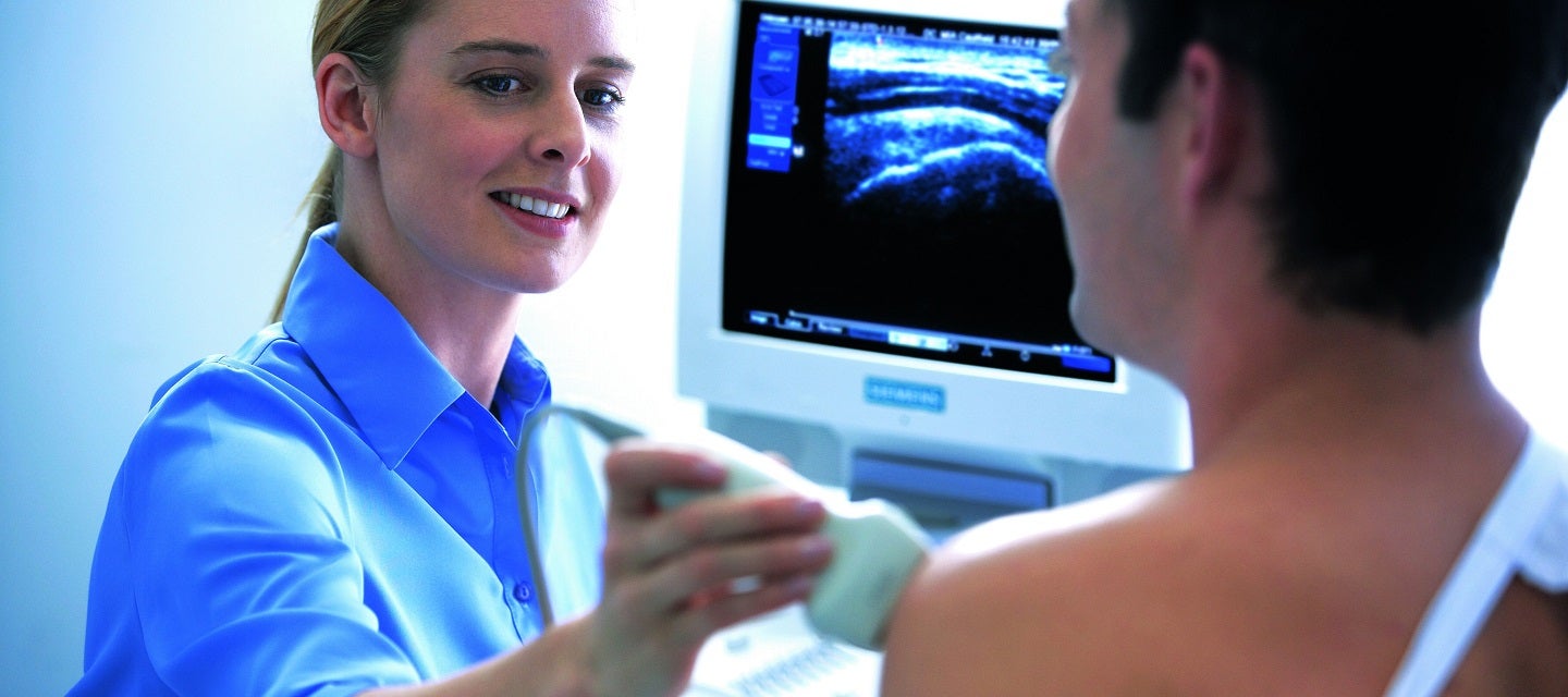
The ultrasound room is darkened and has a narrow bed, beside which there is a machine with a monitor much like a TV or computer screen. The front of the machine looks like a computer keyboard. The part of the machine that is used to produce the ultrasound image is called the "transducer" This is a small plastic square attached to the ultrasound machine by a thick cord.
When you have the ultrasound examination, the Sonographer (technologist who is trained in ultrasound) will spread gel on the area to be examined. This ultrasound gel enables good contact between the transducer and the skin so the best images are obtained. The Sonographer may ask you to move around and/or to hold your breath so they can see the area to be examined clearly.
The Sonographer will watch the monitor during your examination, and may point out fetal anatomy on pregnancy scans.
The Sonographer may ask you to wait in the ultrasound room until they have shown the ultrasound images to the Radiologist. The Radiologist may wish to take some further ultrasound images. This does not mean something is wrong, rather that they may wish to see the moving images of an area.
Who performs the ultrasound? keyboard_arrow_down
The ultrasound examination is performed by a sonographer, a health professional specialised in performing ultrasound examinations. They have a graduate qualification and are fully qualified to perform the examination.
The sonographer performs the examination and provides an interpretation of the images on the screen to a radiologist (medical specialist), who will review the sonographer’s interpretation and discuss the images with them, before providing a report on the findings to your doctor.
Sometimes, it will be necessary for the radiologist to attend the examination, as it may be important to see the images on the screen rather than just the still photographs and discuss your symptoms.
Are there any risks? keyboard_arrow_down
Ultrasound is a safe examination which provides excellent imaging without any significant risk.
What happens after the examination? keyboard_arrow_down
The Radiologist will review the images and produce a written report. Your doctor will receive a copy of the report of your test as soon as is practicable. It is very important that you discuss the results with the doctor whom referred you so that they can explain what the results mean for you.
You will be required to settle your account on the day of the examination.
Related procedures

This information has been reviewed & approved by Dr Ronald Shnier (I-MED Chief Medical Officer).
Related procedures
This information has been reviewed & approved by Dr Ronald Shnier (I-MED Chief Medical Officer).
How much will my examination cost?
Fees for radiology tests can vary and depend on a number of factors. Please make an enquiry with us by phone or email to get a quote for the service you require. ACC co-payments may apply.
We accept all radiology referral forms.
Waikato
Phone: 0800 426 723
Email: Booking.Waikato@i-med.co.nz
Rotorua
Phone: 0800 466 5642
Email: Booking.Rotorua@i-med.co.nz
Bay of Plenty
Phone: 07 544 5993
Email: Booking.bop@i-med.co.nz
Taranaki
Phone: 06 759 4317
Email: Booking.Taranaki@i-med.co.nz