Carotid ultrasound
Carotid ultrasound
What is a carotid ultrasound?
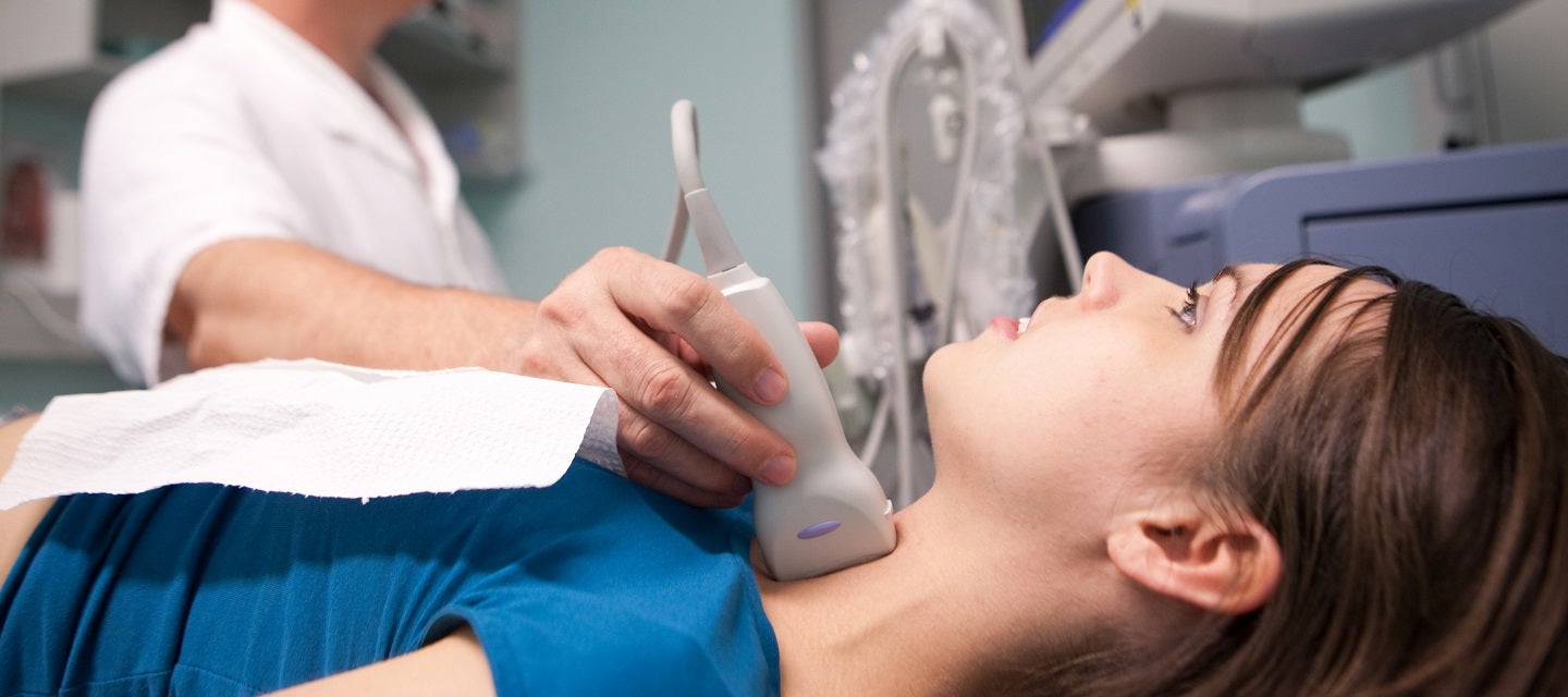
A carotid ultrasound assess the carotid arteries in your neck, that supply blood to your brain.
Ultrasound technology utilises high-frequency sound waves beyond the range of human hearing to create real-time images of the internal structures of the body. In a carotid ultrasound, a transducer emits these sound waves, and when they encounter different tissues or fluids, they produce echoes. The transducer then receives these echoes and sends them to a computer, which processes the information to generate detailed images.
This non-invasive test helps in evaluating the risk of vascular conditions. Your doctor may ask you to have a carotid ultrasound for a number of reasons; including prior to cardiac surgery, after unusual "turns" and after a suspected stroke.
How much will my examination cost?
Fees for radiology tests can vary and depend on a number of factors. Please make an enquiry with us by phone or email to get a quote for the service you require. ACC co-payments may apply.
We accept all radiology referral forms.
Waikato
Phone: 0800 426 723
Email: Booking.Waikato@i-med.co.nz
Rotorua
Phone: 0800 466 5642
Email: Booking.Rotorua@i-med.co.nz
Bay of Plenty
Phone: 07 544 5993
Email: Booking.bop@i-med.co.nz
Taranaki
Phone: 06 759 4317
Email: Booking.Taranaki@i-med.co.nz
How do I prepare for a carotid ultrasound? keyboard_arrow_down
There is very little preparation required.
It is advisable to wear an open-neck shirt or top and not a high necked or collared top, to make it easier to access the vessels of the neck.
Please bring any relevant x-rays or scans.
Who performs the scan? keyboard_arrow_down
The scan will be performed by the Sonographer (Technologist trained specifically in ultrasound).
What happens during the ultrasound? keyboard_arrow_down
You will be asked to lie on a bed and bare your neck. Gel will be spread on your neck, which will allow us to obtain high quality images. A small towel will be used to protect your clothes.
When the Sonographer performs this scan, they will be using colour and pulsed Doppler, which means you will see different colours on the TV monitor and hear different sounds.
The Sonographer may tap you on the side of your head gently during the exam; this is to help identify different vessels.
You will be asked not to talk when the transducer is on your neck, as talking will make the vessels move.
How long does the scan take? keyboard_arrow_down
The procedure takes 30-40 minutes.
What happens after the scan? keyboard_arrow_down
The Radiologist will review the ultrasound images and provide a written report to your doctor.
Related procedures
This information has been reviewed & approved by Dr Ronald Shnier (I-MED Chief Medical Officer).
Related procedures
This information has been reviewed & approved by Dr Ronald Shnier (I-MED Chief Medical Officer).
How much will my examination cost?
Fees for radiology tests can vary and depend on a number of factors. Please make an enquiry with us by phone or email to get a quote for the service you require. ACC co-payments may apply.
We accept all radiology referral forms.
Waikato
Phone: 0800 426 723
Email: Booking.Waikato@i-med.co.nz
Rotorua
Phone: 0800 466 5642
Email: Booking.Rotorua@i-med.co.nz
Bay of Plenty
Phone: 07 544 5993
Email: Booking.bop@i-med.co.nz
Taranaki
Phone: 06 759 4317
Email: Booking.Taranaki@i-med.co.nz