Duplex doppler venous ultrasound
Duplex doppler venous ultrasound
What is a duplex doppler venous ultrasound?
A duplex doppler venous scan utilises ultrasound technology to assess the blood flow in the veins of the legs. This test combines two important aspects:
1. Duplex Ultrasound: This involves using ultrasound to create detailed, real-time images of the leg veins, allowing healthcare professionals to visualise the structure of the veins, identify abnormalities, and assess overall venous anatomy.
2. Doppler Ultrasound: The Doppler component assesses the direction and speed of blood flow within the veins. It provides information on the presence of any obstructions, abnormalities, or conditions affecting the normal flow of blood through the veins.
A duplex doppler venous leg scan provides comprehensive information about both the anatomy and blood flow in the leg veins. This diagnostic tool is commonly employed to identify conditions such as deep vein thrombosis (DVT), venous insufficiency, and other vascular issues in the lower extremities.
How much will my examination cost?
Fees for radiology tests can vary and depend on a number of factors. Please make an enquiry with us by phone or email to get a quote for the service you require. ACC co-payments may apply.
We accept all radiology referral forms.
Waikato
Phone: 0800 426 723
Email: Booking.Waikato@i-med.co.nz
Rotorua
Phone: 0800 466 5642
Email: Booking.Rotorua@i-med.co.nz
Bay of Plenty
Phone: 07 544 5993
Email: Booking.bop@i-med.co.nz
Taranaki
Phone: 06 759 4317
Email: Booking.Taranaki@i-med.co.nz
How do I prepare for the scan? keyboard_arrow_down
No special preparation is required for this examination. Please bring your referral form from the doctor or specialist.
Who performs the scan? keyboard_arrow_down
The scan will be performed by the Sonographer (Technologist trained specifically in ultrasound).
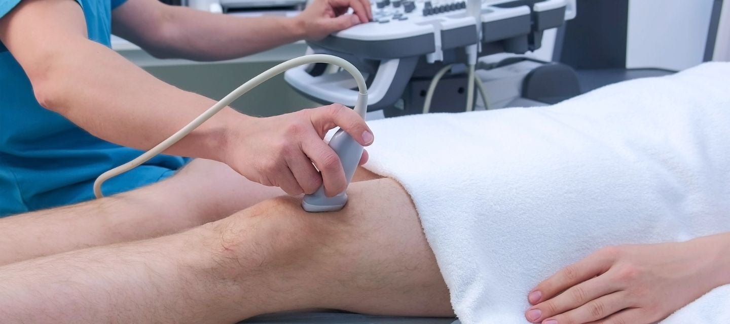
What happens during the scan? keyboard_arrow_down
You may be asked to change into a cotton gown and lie on a narrow bed or remove your trousers. The Sonographer will spread gel on the area to be examined. This will allow us to obtain high quality images of the veins from your groin to your ankle. The Sonographer will ask you to lie on your back initially and then turn onto your stomach about halfway through the examination to examine the veins at the back of your legs.
This will be done with you in a standing or semi-upright position. You will feel pressure as the sonographer assesses the compressibility of you veins and squeezing of your calf muscle to show blood movement.
How long does the scan take? keyboard_arrow_down
The procedure takes 20-30 minutes per leg.
What happens after the scan? keyboard_arrow_down
The Radiologist will review the ultrasound images and provide a written report to your doctor.
Related procedures

This information has been reviewed & approved by Dr Ronald Shnier (I-MED Chief Medical Officer).
Related procedures
This information has been reviewed & approved by Dr Ronald Shnier (I-MED Chief Medical Officer).
How much will my examination cost?
Fees for radiology tests can vary and depend on a number of factors. Please make an enquiry with us by phone or email to get a quote for the service you require. ACC co-payments may apply.
We accept all radiology referral forms.
Waikato
Phone: 0800 426 723
Email: Booking.Waikato@i-med.co.nz
Rotorua
Phone: 0800 466 5642
Email: Booking.Rotorua@i-med.co.nz
Bay of Plenty
Phone: 07 544 5993
Email: Booking.bop@i-med.co.nz
Taranaki
Phone: 06 759 4317
Email: Booking.Taranaki@i-med.co.nz