X-ray
X-ray
What is an x-ray?
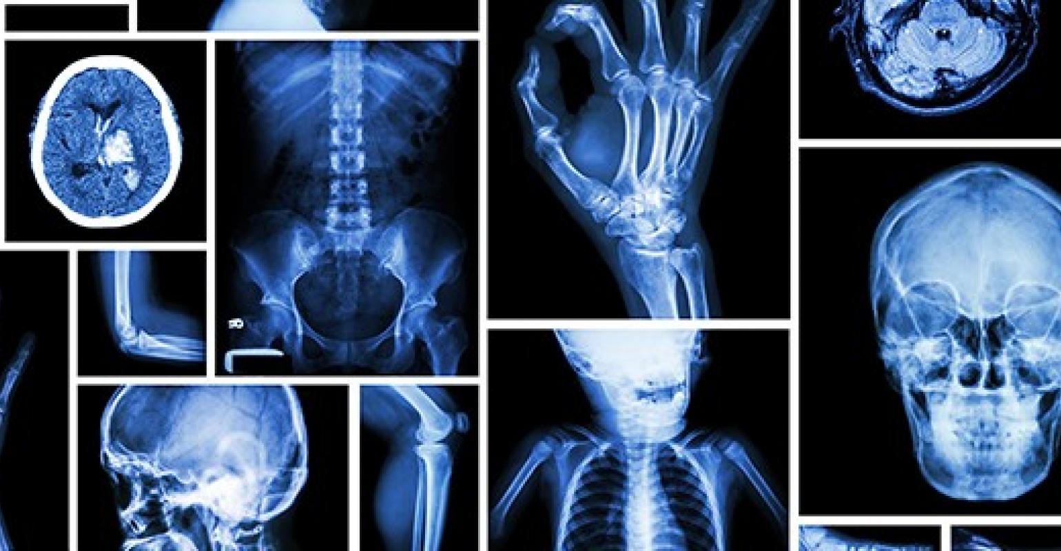
An x-ray is the oldest and most frequently used form of medical imaging.
Images are created by passing controlled amounts of radiation through the body. The radiation exiting from the body strikes a digital x-ray plate capturing this energy, the image is detected with a light camera and an image created with the aid of computer software.
X-ray imaging is the fastest and easiest way for a doctor to view and assess broken bones and other skeletal abnormalities. At least two digital image plates are taken of a bone, and often three digital image plates if the problem is around a joint (knee, elbow, or wrist). X-rays are also commonly taken of the Chest and Abdomen.
How much will my examination cost?
Fees for radiology tests can vary and depend on a number of factors. Please make an enquiry with us by phone or email to get a quote for the service you require. ACC co-payments may apply.
We accept all radiology referral forms.
Waikato
Phone: 0800 426 723
Email: Booking.Waikato@i-med.co.nz
Rotorua
Phone: 0800 466 5642
Email: Booking.Rotorua@i-med.co.nz
Bay of Plenty
Phone: 07 544 5993
Email: Booking.bop@i-med.co.nz
Taranaki
Phone: 06 759 4317
Email: Booking.Taranaki@i-med.co.nz
How do I prepare for an x-ray? keyboard_arrow_down
You may be asked to change into a gown before your examination, but wearing light clothing which is free of zips and buttons can often be x-rayed through.
You will also be asked to remove jewelry, glasses, and any metal objects that could obscure the images, since those show up on x-ray.
Women should always inform their doctor or the medical imaging technologist if there is any possibility that they are pregnant.
How is an x-ray performed? keyboard_arrow_down
The technologist (also known as the 'radiographer') positions the patient, places a flat image plate under the table or directly under the area of the body to be imaged. Depending on the body site, imaging can be performed standing, sitting or lying down.
The technologist goes to a small adjacent room and asks the patient to hold very still without breathing for a few seconds.
The radiographic equipment is activated, sending a beam of x-rays through the body to expose the digital image plate. The technologist then repositions the patient for another view, and the process is repeated. When the x-rays are completed you will be asked to wait a short time until the technologist reviews the images to determine if more are needed.
What will I experience during the x-ray examination? keyboard_arrow_down
In most cases, x-ray imaging is painless. Sometimes, to get a clear image of an injury such as a possible fracture, you may be asked to hold an uncomfortable position for a short time. Any movement could blur the image and make it necessary to repeat the procedure to get a useful, clear picture.
How long does an x-ray take? keyboard_arrow_down
Usually, an x-ray examination takes less than 15 minutes.
Who interprets the x-ray results and how do I get them? keyboard_arrow_down
A radiologist, who is a specialist doctor experienced in bone x-ray and other radiology examinations, will analyse the images and send a written report with his or her interpretation to the patient's referring doctor.
Related procedures

This information has been reviewed & approved by Dr Ronald Shnier (I-MED Chief Medical Officer).
Related procedures
This information has been reviewed & approved by Dr Ronald Shnier (I-MED Chief Medical Officer).
How much will my examination cost?
Fees for radiology tests can vary and depend on a number of factors. Please make an enquiry with us by phone or email to get a quote for the service you require. ACC co-payments may apply.
We accept all radiology referral forms.
Waikato
Phone: 0800 426 723
Email: Booking.Waikato@i-med.co.nz
Rotorua
Phone: 0800 466 5642
Email: Booking.Rotorua@i-med.co.nz
Bay of Plenty
Phone: 07 544 5993
Email: Booking.bop@i-med.co.nz
Taranaki
Phone: 06 759 4317
Email: Booking.Taranaki@i-med.co.nz