PET-CT
PET-CT
What is a PET-CT scan?
There are two parts to a PET-CT scan, both are performed on the same scanner at the same time.
Positron emission tomography, known as PET, is a nuclear medicine imaging test in which a small amount of liquid radioactive material is injected into your body to provide functional information about the body. The radioactive substance most used in PET scanning is a simple sugar (like glucose) called FDG, which stands for “fluorodeoxyglucose”. It is injected into your bloodstream and accumulates in your body where it gives off energy in the form of gamma rays. These are detected by the PET scanner and a computer converts the signals into detailed pictures or images showing how tissue and organs are working. If you are having an FDG PET, your sugar metabolism (how sugar is used by your body) is imaged. More information on the type of radioactive tracers is provided below.
CT (Computed tomography) imaging uses x-ray equipment to create detailed images of slices of the anatomy/structures inside of your body.
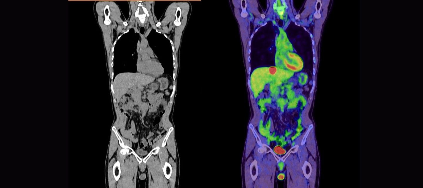
Both PET and CT scan information is displayed in the form of images and then combined or fused to form the PET-CT.
The PET-CT combination allows any abnormality on the PET scan to be precisely located within the body, allowing for accurate diagnosis of a variety of conditions, including many types of cancers, heart and other diseases.
How much will my examination cost?
Fees for radiology tests can vary and depend on a number of factors. Please make an enquiry with us by phone or email to get a quote for the service you require. ACC co-payments may apply.
We accept all radiology referral forms.
Waikato
Phone: 0800 426 723
Email: Booking.Waikato@i-med.co.nz
Rotorua
Phone: 0800 466 5642
Email: Booking.Rotorua@i-med.co.nz
Bay of Plenty
Phone: 07 544 5993
Email: Booking.bop@i-med.co.nz
Taranaki
Phone: 06 759 4317
Email: Booking.Taranaki@i-med.co.nz
How do I prepare for a PET scan? keyboard_arrow_down
Our team will inform you of the exact preparation that you will need for the scan. It is very important to follow the preparation instructions, failure to do so could result in the scan being postponed or cancelled.
Fasting
Generally, you will be asked not to eat or drink anything for several hours before the PET scan because this may alter your sugar metabolism and may affect the quality of the images or pictures. Drinking water is usually acceptable. If you are diabetic, you will be provided with specific instructions and may need to stop taking some diabetic medications before having the scan.
Pregnancy and breastfeeding
It is important that you let our staff know if you are (or think you could be) pregnant or are breast feeding. As a rule, administration of any drug to a pregnant woman, including contrast media, needs to be carefully considered.
This study may not be suitable for pregnant women because of the radiation dose to the growing foetus. Please discuss this with your doctor.
Women who are breastfeeding and people who are the primary or sole carer for small children may need to make special preparations for after the test, to stop breastfeeding for a short time, and to avoid close contact with young children. This is due to the small amount of radioactivity your body may release for a while after the test. Talk to your referring doctor or our clinic staff for details.
Previous scans
Bring with you to your appointment any previous X-ray or radiology images you have, as comparison with these by the nuclear medicine physician (a specialist doctor), who looks at and interprets your PET scan, can be very helpful.
Clothing
You need to wear comfortable, loose clothing and will generally be changed into a hospital gown. It is important that you are not wearing metal, including jewellery, watches, zips and bra hooks as these can affect the quality of the images produced.
What is 18F-FDG? keyboard_arrow_down
18F-FDG is a radioactive sugar that is absorbed into high glucose-using cells such as the brain, heart, kidneys and if present in the body, active tumour cells. 18F-FDG is comprised of two components; a radioactive isotope of fluorine (18F) and a glucose (sugar) base tracer of fluorodeoxyglucose (FDG). Adverse reactions are extremely rare and the radiation exposure is comparable to a conventional CT scan.
What happens during a PET/CT scan? keyboard_arrow_down
A nuclear medicine technologist (a healthcare professional trained to handle isotopes) will administer an individually tailored injection into a vein (usually in your arm). Following this injection, you will be asked to rest quietly in your own uptake room - feel free to bring a book or tablet to occupy yourself as relatives and friends will be asked to wait in the waiting area.
You will then be moved to the scanning room and positioned on the PET-CT scanning bed. It is important to remain as still as possible during the scan as movement can result in reduced image quality and the images may be blurry. Therefore, if you are uncomfortable after being positioned on the bed please tell the technologist.
The intravenous line will be removed before you leave.
How long does the scan take? keyboard_arrow_down
The scan time takes between 15-40 minutes (the exact scan time will be given before we start). From arrival to departure, you will be with us approximately 3 hours.
What to expect afterwards keyboard_arrow_down
The technologist will tell you if there are any special instructions you need to follow after the scan. In most cases you will be able to leave straight away, then eat and drink as normal. The isotope loses its radioactivity over time and will pass naturally out of your body. To assist passing the isotope out of your body, we encourage you to drink plenty of liquid and empty your bladder regularly. The amount of radiation in your body after the scan will be very small, if you have any concerns about exposure to radiation, please discuss this with one of our team.
How do I get my results? keyboard_arrow_down
The PET-CT images will be electronically sent following the scan. A written report will be sent to the doctor who has requested your scan (usually within 48 hours), please inform us if you would like a copy of the report to be sent to your GP. Your doctor will be able to discuss the results with you at your next appointment.
Types of radioactive tracers used in PET imaging keyboard_arrow_down
18F-FDG
18F-FDG is a radioactive sugar that is absorbed into high-glucose-using cells. 18F-FDG is comprised of two components; a radioactive isotope of fluorine (18F) and a glucose (sugar) base tracer of fluorodeoxyglucose (FDG).
18F-FDG PET CT is commonly used in the evaluation and management of many types of cancer including tumour diagnosis, staging, restaging, treatment monitoring, and radiation therapy planning. It is also used in the evaluation of myocardial viability, sarcoidosis, infection and assessment of neurological conditions including epilepsy and dementia.
18F-NaF
18F-NaF is bone-seeking tracer used to image skeletal abnormalities. 18F-NaF is comprised of two components; a radioactive isotope of fluorine (18F) and base tracer of Sodium Fluoride (NaF). 18F-NaF PET CT is commonly used in the evaluation and management for back pain, unexplained bone pain, abnormal radiographic or laboratory findings, osteomyelitis, trauma, inflammatory and degenerative arthritis, avascular necrosis, osteonecrosis of the mandible, condylar hyperplasia, metabolic bone disease, Paget’s disease, NAI, bone graft viability, complications of prosthetic joints, reflex sympathetic dystrophy and distribution of osteoblastic activity prior to administration of therapeutic radiopharmaceuticals for treating bone pain.
18F-PSMA
18F-PSMA is a prostate-specific membrane antigen (PSMA) and a recognized target for imaging prostate cancer. 18F-PSMA is comprised of two components; a radioactive isotope of fluorine (18F) and base tracer of DCFPyL (PSMA or PSR).
18F-PSMA PET CT is used for staging and re-staging prostate cancer. 18F-PSMA is the newest molecular imaging technique in New Zealand, and we are proud to offer this service at Midland PET CT, part of the I-MED Radiology Network.
Adverse reactions to the above are extremely rare, and the radiation exposure is comparable to a conventional CT scan. If you have any additional questions, please feel free to talk about them with your doctor or one of our team before the scan.
Related procedures

This information has been reviewed & approved by Dr Ronald Shnier (I-MED Chief Medical Officer).
Related procedures
This information has been reviewed & approved by Dr Ronald Shnier (I-MED Chief Medical Officer).
How much will my examination cost?
Fees for radiology tests can vary and depend on a number of factors. Please make an enquiry with us by phone or email to get a quote for the service you require. ACC co-payments may apply.
We accept all radiology referral forms.
Waikato
Phone: 0800 426 723
Email: Booking.Waikato@i-med.co.nz
Rotorua
Phone: 0800 466 5642
Email: Booking.Rotorua@i-med.co.nz
Bay of Plenty
Phone: 07 544 5993
Email: Booking.bop@i-med.co.nz
Taranaki
Phone: 06 759 4317
Email: Booking.Taranaki@i-med.co.nz