DVT ultrasound
DVT ultrasound
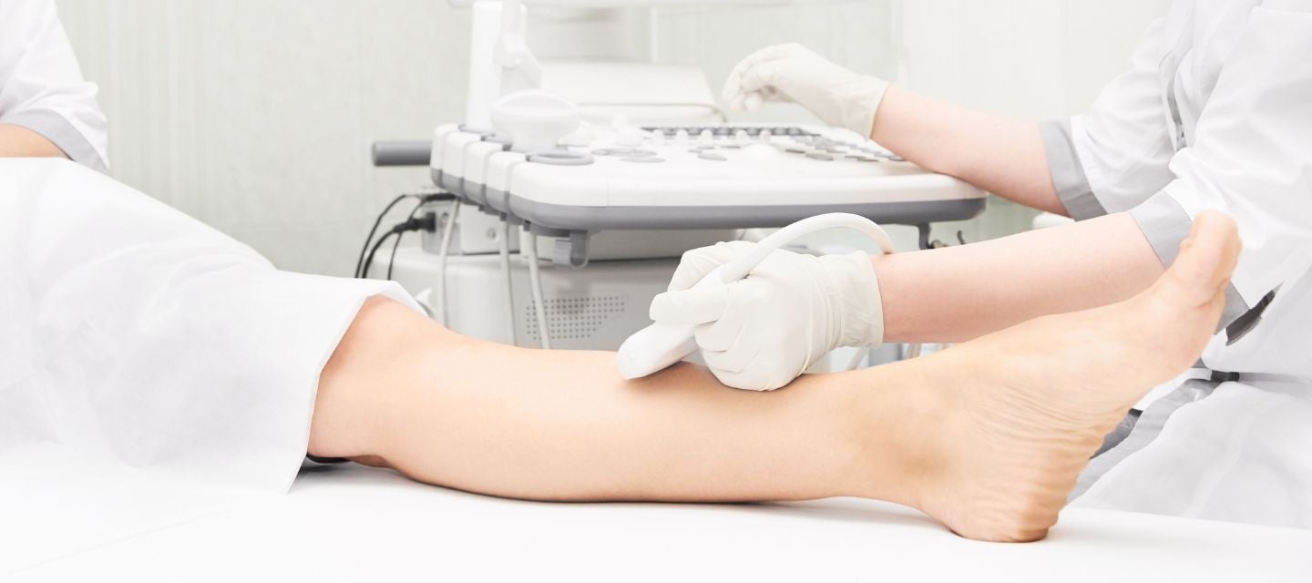
What is a DVT ultrasound?
A deep vein thrombosis (DVT) ultrasound is a medical imaging test that uses ultrasound technology to non-invasively assess and visualise blood flow in the deep veins of the upper and lower extremities, aiding in the diagnosis of potential blood clots.
Your doctor may recommend this scan to exclude clots in the veins, especially if you experience symptoms such as a swollen or painful leg and have a history of trauma, surgery, or long-distance flights.
How much will my examination cost?
Fees for radiology tests can vary and depend on a number of factors. Please make an enquiry with us by phone or email to get a quote for the service you require. ACC co-payments may apply.
We accept all radiology referral forms.
Waikato
Phone: 0800 426 723
Email: Booking.Waikato@i-med.co.nz
Rotorua
Phone: 0800 466 5642
Email: Booking.Rotorua@i-med.co.nz
Bay of Plenty
Phone: 07 544 5993
Email: Booking.bop@i-med.co.nz
Taranaki
Phone: 06 759 4317
Email: Booking.Taranaki@i-med.co.nz
How do I prepare for the scan? keyboard_arrow_down
No special preparation is required for this examination. Please bring your referral form from the doctor or specialist.
Who performs the ultrasound? keyboard_arrow_down
The ultrasound examination is performed by a sonographer, a health professional specialised in performing ultrasound examinations. They have a graduate qualification and are fully qualified to perform the examination.
The sonographer performs the examination and provides an interpretation of the images on the screen to a radiologist (medical specialist), who will review the sonographer’s interpretation and discuss the images with them, before providing a report on the findings to your doctor.
Sometimes, it will be necessary for the radiologist to attend the examination, as it may be important to see the images on the screen rather than just the still photographs and discuss your symptoms.
What happens during the scan? keyboard_arrow_down
You will be asked to lie on a narrow bed. The Sonographer will spread gel on the area to be examined usually from the groin to the ankle. This will allow us to obtain high quality images. The Sonographer will ask you to lie on your back initially and then possibly turn onto your stomach about halfway through the examination to examine the veins at the back of your legs.
How long does the scan take? keyboard_arrow_down
This procedure takes 20-30 minutes.
What happens after the scan? keyboard_arrow_down
The Radiologist will review the ultrasound images and provide a written to your Doctor. If there is a clot, we will ring your doctor while you are still here to see what the next step in your care will be.
Related procedures

This information has been reviewed & approved by Dr Ronald Shnier (I-MED Chief Medical Officer).
Related procedures
This information has been reviewed & approved by Dr Ronald Shnier (I-MED Chief Medical Officer).
How much will my examination cost?
Fees for radiology tests can vary and depend on a number of factors. Please make an enquiry with us by phone or email to get a quote for the service you require. ACC co-payments may apply.
We accept all radiology referral forms.
Waikato
Phone: 0800 426 723
Email: Booking.Waikato@i-med.co.nz
Rotorua
Phone: 0800 466 5642
Email: Booking.Rotorua@i-med.co.nz
Bay of Plenty
Phone: 07 544 5993
Email: Booking.bop@i-med.co.nz
Taranaki
Phone: 06 759 4317
Email: Booking.Taranaki@i-med.co.nz