Vascular ultrasound
Vascular ultrasound
What is a vascular ultrasound?
The high spec ultrasound machine utilises Colour and Pulsed Doppler to assess the veins and arteries, to measure the speed and direction of the blood flow.
Some of the indications of vascular ultrasound are given below.
Stroke:
Many strokes are caused by abnormal blood flow in the neck. These are easily examined with colour Doppler ultrasound.
High Blood Pressure/Kidneys:
The blood flow to, and within the kidney can be assessed to look for a cause for high blood pressure.
Arterial Bypass Grafts:
Regular (6-12 monthly) examination of these grafts is essential. The detection of early graft failure can allow the graft to be revised before it fails.
Varicose Veins:
These are usually done on referral from a surgeon. The type of varicose vein abnormalities can be mapped and marked pre-operatively to help your Surgeon.
How much will my examination cost?
Fees for radiology tests can vary and depend on a number of factors. Please make an enquiry with us by phone or email to get a quote for the service you require. ACC co-payments may apply.
We accept all radiology referral forms.
Waikato
Phone: 0800 426 723
Email: Booking.Waikato@i-med.co.nz
Rotorua
Phone: 0800 466 5642
Email: Booking.Rotorua@i-med.co.nz
Bay of Plenty
Phone: 07 544 5993
Email: Booking.bop@i-med.co.nz
Taranaki
Phone: 06 759 4317
Email: Booking.Taranaki@i-med.co.nz
How do I prepare for the scan? keyboard_arrow_down
No special preparation is required for this examination. Please bring your referral form from the doctor or specialist.
How long does the scan take? keyboard_arrow_down
On average the scan takes 30-40 minutes.
Who performs the scan? keyboard_arrow_down
The scan will be performed by the Sonographer (Technologist trained specifically in ultrasound).
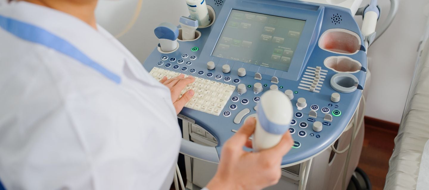
What happens during a vascular ultrasound? keyboard_arrow_down
During the procedure, a handheld device (called a transducer) emits high-frequency sound waves and acts as both a transmitter and a receiver. The transducer is gently moved across the skin in the area being examined, sending sound waves into the body and collecting the echoes that bounce back. These echoes are then converted into real-time images on a computer screen, allowing healthcare professionals to visualise the blood vessels and assess blood flow.
To ensure efficient sound wave transmission and prevent air pockets that can interfere with imaging, a water-based gel is applied to the skin at the examination site. This gel helps create a secure and smooth interface between the transducer and the skin, facilitating the transmission of sound waves and enhancing the quality of the ultrasound images. The use of gel also helps minimise discomfort for the patient during the procedure.
What happens after the scan? keyboard_arrow_down
The Radiologist will review the ultrasound images and provide a written report to your doctor.
Related procedures
This information has been reviewed & approved by Dr Ronald Shnier (I-MED Chief Medical Officer).
Related procedures
This information has been reviewed & approved by Dr Ronald Shnier (I-MED Chief Medical Officer).
How much will my examination cost?
Fees for radiology tests can vary and depend on a number of factors. Please make an enquiry with us by phone or email to get a quote for the service you require. ACC co-payments may apply.
We accept all radiology referral forms.
Waikato
Phone: 0800 426 723
Email: Booking.Waikato@i-med.co.nz
Rotorua
Phone: 0800 466 5642
Email: Booking.Rotorua@i-med.co.nz
Bay of Plenty
Phone: 07 544 5993
Email: Booking.bop@i-med.co.nz
Taranaki
Phone: 06 759 4317
Email: Booking.Taranaki@i-med.co.nz