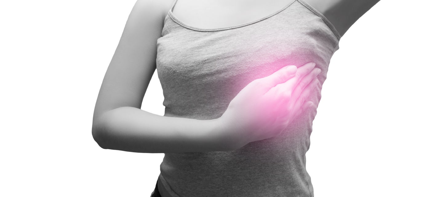
Your complete guide to breast care
Explore our trusted breast care resources to help you stay informed, proactive, and confident in making decisions about your health. When yo ...
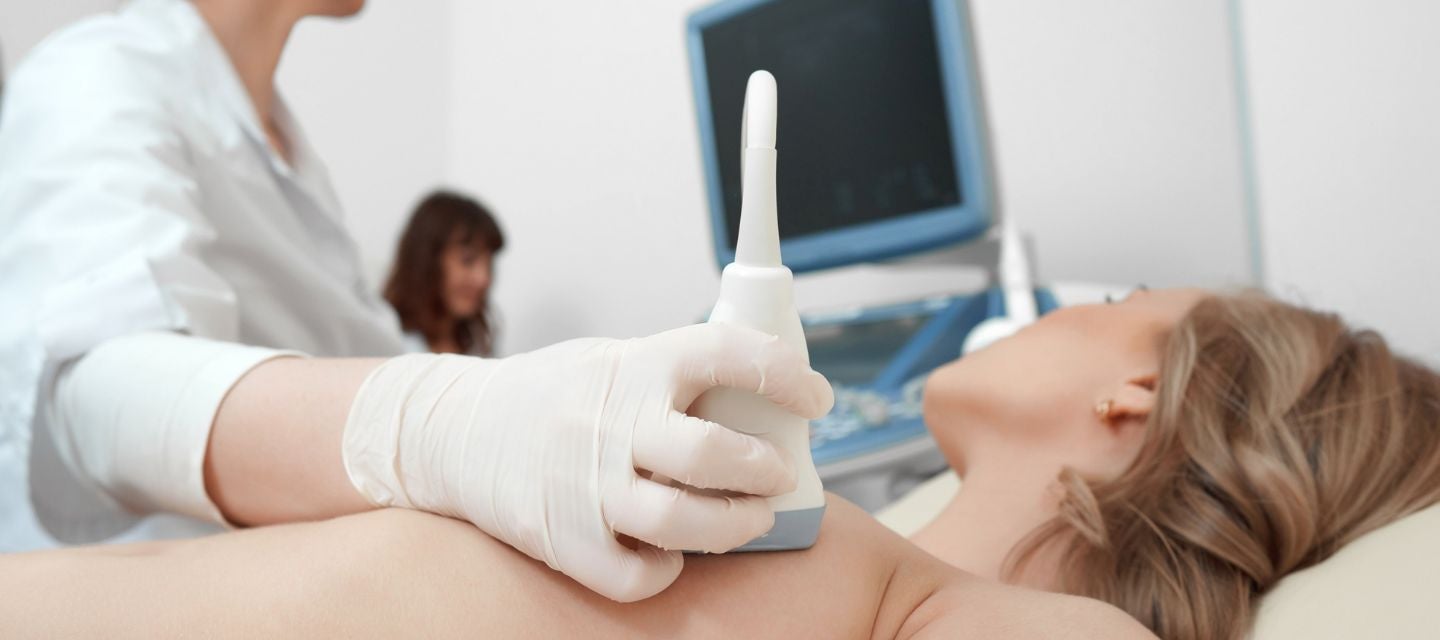
Ultrasound of the breast helps to distinguish fluid filled lumps in the breast (cysts), from solid lumps which may be cancerous or benign (non-cancerous). It is often useful to examine the breasts of younger women because the breast tissue is much denser than it is in older women, and this density can make it harder to detect an abnormality if a mammogram is performed.
Ultrasound is also used to diagnose problems such as complications from mastitis (an infection that occurs most often during breastfeeding), to assess abnormal nipple discharge, problems with breast implants, and to guide the placement of a needle during biopsies.
Fees for radiology tests can vary and depend on a number of factors. Please make an enquiry with us by phone or email to get a quote for the service you require. ACC co-payments may apply.
We accept all radiology referral forms.
Waikato
Phone: 0800 426 723
Email: Booking.Waikato@i-med.co.nz
Rotorua
Phone: 0800 466 5642
Email: Booking.Rotorua@i-med.co.nz
Bay of Plenty
Phone: 07 544 5993
Email: Booking.bop@i-med.co.nz
Taranaki
Phone: 06 759 4317
Email: Booking.Taranaki@i-med.co.nz
No preparation is necessary for this examination.
It is advisable to wear a two piece outfit so that only your top has to be removed to provide access to the breast area.
You will be asked to lie on a bed and one breast at a time will be examined.
Gel (a water-based jelly) is applied to the skin and an ultrasound probe (called a transducer) is placed on the breast and gently moved around the breast to examine the breast tissue. This produces pictures on a screen of the tissues inside your breast, in the same way that a pregnancy ultrasound scan is performed.
Examination of the armpit (or axilla) may also be undertaken to assess for any enlarged lymph glands (or nodes – a lump or swelling).
The examination takes between 15-30 minutes. Sometimes the sonographer will ask you to wait and have the images checked by the radiologist (specialist doctor).
The Radiologist will review the images and provide a written report to your referring doctor. Please settle your account on the day of the examination.

This information has been reviewed & approved by Dr Ronald Shnier (I-MED Chief Medical Officer).
This information has been reviewed & approved by Dr Ronald Shnier (I-MED Chief Medical Officer).
Fees for radiology tests can vary and depend on a number of factors. Please make an enquiry with us by phone or email to get a quote for the service you require. ACC co-payments may apply.
We accept all radiology referral forms.
Waikato
Phone: 0800 426 723
Email: Booking.Waikato@i-med.co.nz
Rotorua
Phone: 0800 466 5642
Email: Booking.Rotorua@i-med.co.nz
Bay of Plenty
Phone: 07 544 5993
Email: Booking.bop@i-med.co.nz
Taranaki
Phone: 06 759 4317
Email: Booking.Taranaki@i-med.co.nz