Musculoskeletal ultrasound
Musculoskeletal ultrasound
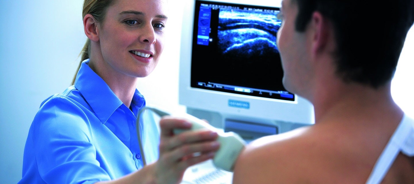
What is a musculoskeletal ultrasound?
A musculoskeletal ultrasound is a medical imaging technique that uses high-frequency sound waves to create real-time, detailed images of the muscles, tendons, ligaments, joints, and other soft tissues within the musculoskeletal system.
This non-invasive diagnostic tool allows healthcare professionals to visualise the internal structures of the musculoskeletal system and assess for abnormalities, injuries, inflammation, or other conditions affecting the bones and soft tissues.
Musculoskeletal ultrasound is commonly used to diagnose and monitor conditions such as tendonitis, ligament injuries, joint inflammation, and other musculoskeletal disorders. It provides valuable information for treatment planning and can be performed in conjunction with other imaging modalities to offer a comprehensive assessment of musculoskeletal health.
How much will my examination cost?
Fees for radiology tests can vary and depend on a number of factors. Please make an enquiry with us by phone or email to get a quote for the service you require. ACC co-payments may apply.
We accept all radiology referral forms.
Waikato
Phone: 0800 426 723
Email: Booking.Waikato@i-med.co.nz
Rotorua
Phone: 0800 466 5642
Email: Booking.Rotorua@i-med.co.nz
Bay of Plenty
Phone: 07 544 5993
Email: Booking.bop@i-med.co.nz
Taranaki
Phone: 06 759 4317
Email: Booking.Taranaki@i-med.co.nz
How do I prepare for a musculoskeletal ultrasound? keyboard_arrow_down
There is no special preparation for this scan.
Who performs the scan? keyboard_arrow_down
The scan will be performed by the Sonographer (Technologist trained specifically in ultrasound).
What are the indications for a musculoskeletal ultrasound? keyboard_arrow_down
While ultrasound will not show every soft tissue abnormality, the following are standard applications for performing the scan
Muscle ultrasound: Muscle injury, large tears and haematomas (bruises).
Tendon: These connect muscle to bone. Tendons in many parts of the body are readily visible with ultrasound, in particular the shoulder, ankle and Achilles tendon.
Bursae: These are sac-like structures filled with fluid found close to joints. Superficial bursae are found between bone and skin. Deep bursae separate a joint from overlying tendons and ligaments. A swollen bursa can be detected with ultrasound, most commonly seen around the shoulder, knee joints and hip.
Ligaments: These connect bone to bone. Some ligaments can be demonstrated with ultrasound. Others require Magnetic Resonance Imaging (MRI).
Foreign bodies: Not all foreign bodies will be visible using x-rays. Most will be visible with ultrasound. The location of the foreign body and damage to any surrounding structures is readily seen. A skin mark can be applied so the foreign body can be removed at a later time.
How long will the ultrasound take? keyboard_arrow_down
Most scans take between 15 and 30 minutes. In some cases, the normal (asymptomatic) side may also be examined.
What happens after the examination? keyboard_arrow_down
The Radiologist will review the images and produce a written report. Your doctor will receive a copy of the report of your test as soon as is practicable. It is very important that you discuss the results with the doctor whom referred you so that they can explain what the results mean for you.
You will be required to settle your account on the day of the examination.
Related procedures

This information has been reviewed & approved by Dr Ronald Shnier (I-MED Chief Medical Officer).
Related procedures
This information has been reviewed & approved by Dr Ronald Shnier (I-MED Chief Medical Officer).
How much will my examination cost?
Fees for radiology tests can vary and depend on a number of factors. Please make an enquiry with us by phone or email to get a quote for the service you require. ACC co-payments may apply.
We accept all radiology referral forms.
Waikato
Phone: 0800 426 723
Email: Booking.Waikato@i-med.co.nz
Rotorua
Phone: 0800 466 5642
Email: Booking.Rotorua@i-med.co.nz
Bay of Plenty
Phone: 07 544 5993
Email: Booking.bop@i-med.co.nz
Taranaki
Phone: 06 759 4317
Email: Booking.Taranaki@i-med.co.nz