Pelvis/transvaginal ultrasound
Pelvis/transvaginal ultrasound
What is an ultrasound of the female pelvis?
A pelvis or transvaginal ultrasound is a medical imaging scan used to examine the pelvic organs in females. This test is recommended by doctors for various reasons, such as pelvic pain, menstrual problems, and post-menopausal bleeding. The ultrasound helps provide detailed images of the reproductive organs, assisting healthcare professionals in diagnosing and monitoring conditions related to the pelvic region.
Patients undergoing a pelvic ultrasound typically require an transvaginal scan, especially for optimal imaging of the uterus's anatomy, including the endometrium, and the ovaries.
The ultrasound examination can be conducted in two ways:
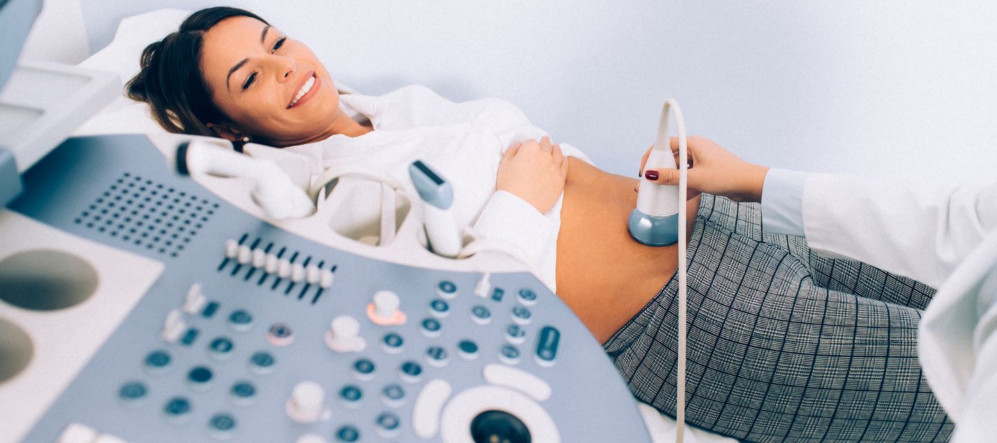
- Trans-abdominal: The ultrasound probe is placed on the lower abdomen and gently moved over the skin to capture images of the pelvic organs.
- transvaginal: A tampon-sized probe is inserted into the vagina to obtain detailed images of the uterus and ovaries. This method is particularly useful for examining the lining of the uterus (endometrium).
How much will my examination cost?
Fees for radiology tests can vary and depend on a number of factors. Please make an enquiry with us by phone or email to get a quote for the service you require. ACC co-payments may apply.
We accept all radiology referral forms.
Waikato
Phone: 0800 426 723
Email: Booking.Waikato@i-med.co.nz
Rotorua
Phone: 0800 466 5642
Email: Booking.Rotorua@i-med.co.nz
Bay of Plenty
Phone: 07 544 5993
Email: Booking.bop@i-med.co.nz
Taranaki
Phone: 06 759 4317
Email: Booking.Taranaki@i-med.co.nz
Who performs the pelvic ultrasound? keyboard_arrow_down
A Sonographer (ultrasound technologist) and/ or Radiologist (Doctor specialising in Radiology) will be involved in your examination. The Sonographer or Radiologist will perform the examination and record the ultrasound. The Radiologist will review these images and provide a written report to your Doctor.
How do I prepare for a pelvis/transvaginal ultrasound? keyboard_arrow_down
You will need to ensure you have a full bladder by drinking 500 ml of water one hour before the scheduled time. The presence of water in the bladder aids in visualising the pelvic organs during the examination.
Can I have the ultrasound if I am bleeding? keyboard_arrow_down
Yes, we can still do this examination if you are bleeding.
If you are wearing a tampon, it will need to be removed. If you are having a period this is not a problem and in some instances it is an advantage when assessing a variety of gynaecological problems.
What happens during the ultrasound? keyboard_arrow_down
The ultrasound pelvis examination can be performed two ways:
A trans-abdominal ultrasound, where the ultrasound probe is placed on your lower abdomen and gently moved over the skin to take the ultrasound images.
An transvaginal ultrasound, where a tampon-sized probe is inserted into the vagina. This method is used to obtain optimum images of the anatomy of the uterus, particularly on the lining of the uterus (the endometrium) and of the ovaries. The transvaginal ultrasound probe is covered in a plastic sheath and covered in ultrasound gel. The transvaginal scan is only used in consultation with the patient.
How long does the examination take? keyboard_arrow_down
A pelvic ultrasound appointment takes 20 - 30 minutes. The transvaginal component, if performed, takes about 10 minutes.
What happens after the scan? keyboard_arrow_down
The Radiologist will review the ultrasound images and provide a written report to your doctor.
Related procedures

This information has been reviewed & approved by Dr Ronald Shnier (I-MED Chief Medical Officer).
Related procedures
This information has been reviewed & approved by Dr Ronald Shnier (I-MED Chief Medical Officer).
How much will my examination cost?
Fees for radiology tests can vary and depend on a number of factors. Please make an enquiry with us by phone or email to get a quote for the service you require. ACC co-payments may apply.
We accept all radiology referral forms.
Waikato
Phone: 0800 426 723
Email: Booking.Waikato@i-med.co.nz
Rotorua
Phone: 0800 466 5642
Email: Booking.Rotorua@i-med.co.nz
Bay of Plenty
Phone: 07 544 5993
Email: Booking.bop@i-med.co.nz
Taranaki
Phone: 06 759 4317
Email: Booking.Taranaki@i-med.co.nz