Pregnancy ultrasound
Pregnancy ultrasound
What is a pregnancy ultrasound?
An ultrasound examination is performed using a smooth, hand held device called a transducer (camera) that moves across the body with a sliding and rotating action. The transducer transmits high-frequency sound waves into your body. The sound waves are then reflected from the different tissues in different ways and converted to electrical impulses, which are used to produce a moving image onto the screen.
Ultrasound is widely used in medical care during pregnancy. It is an ideal examination to look at the baby as it grows throughout the various stages of pregnancy, and is a wonderful opportunity to meet your forming baby.
Routine pregnancy scans are performed in at 12-13 weeks gestation and again at 20 weeks. Your LMC may recommend a scan at other times in your pregnancy to answer a specific question.
How much will my examination cost?
Fees for radiology tests can vary and depend on a number of factors. Please make an enquiry with us by phone or email to get a quote for the service you require. ACC co-payments may apply.
We accept all radiology referral forms.
Waikato
Phone: 0800 426 723
Email: Booking.Waikato@i-med.co.nz
Rotorua
Phone: 0800 466 5642
Email: Booking.Rotorua@i-med.co.nz
Bay of Plenty
Phone: 07 544 5993
Email: Booking.bop@i-med.co.nz
Taranaki
Phone: 06 759 4317
Email: Booking.Taranaki@i-med.co.nz
Why do I need an ultrasound in pregnancy? keyboard_arrow_down
The most common reason to have ultrasound in pregnancy is to determine the age (gestation) of the baby, to confirm its development and well-being, and to make sure the baby is growing normally.
First trimester scans (before 12 weeks gestation) are often performed to assess the health of the forming pregnancy when there is pain or bleeding or to accurately determine gestational age when dates are uncertain. If you are unsure of your dates dating ultrasound is best performed at 8-10 weeks. Early in the pregnancy, we may need to scan with a special transducer placed in the vagina (an endovaginal scan). At the time of your appointment we will assess whether this is required and discuss this with you then.
The Nuchal Translucency assessment is performed between 12 weeks and 13 weeks 6 days. See Nuchal Translucency Scan.
At 20 weeks we will look specifically at the baby’s anatomy (how the baby has been formed), the fluid around the baby, the placenta and assess if the pregnancy is progressing normally. This is the time we can usually determine fetal gender if desired.
How do I prepare for the scan? keyboard_arrow_down
There is very little preparation needed.
If you are in the first 14 weeks of your pregnancy, you may be asked to have a full bladder for your scan. This allows the urine in the bladder to be used as a clear "viewing window" so the baby can be optimally seen. After 14 weeks, having a full bladder when you come for your appointment is not necessary.
For certain types of scan, the gestational age may be very important to ensure the best measurements. Please be patient with the staff making the appointment.
It is a good idea to wear comfortable clothing that gives easy access to your entire abdominal area.
Family and friends are welcome to come with you, however the ultrasound room is small and will only accommodate one extra support person. This will allow your sonographer to focus on producing a thorough examination for you and your baby.
Who performs the scan? keyboard_arrow_down
The scan will be performed by the Sonographer (Technologist trained specifically in ultrasound).
What happens during a pregnancy ultrasound? keyboard_arrow_down
You are asked to lie on an examination couch. The abdomen is exposed and a clear gel is applied to the skin. This can easily be washed off after the examination.
A transducer (a smooth handheld device) is moved gently across the abdomen with a sliding and rotating action.
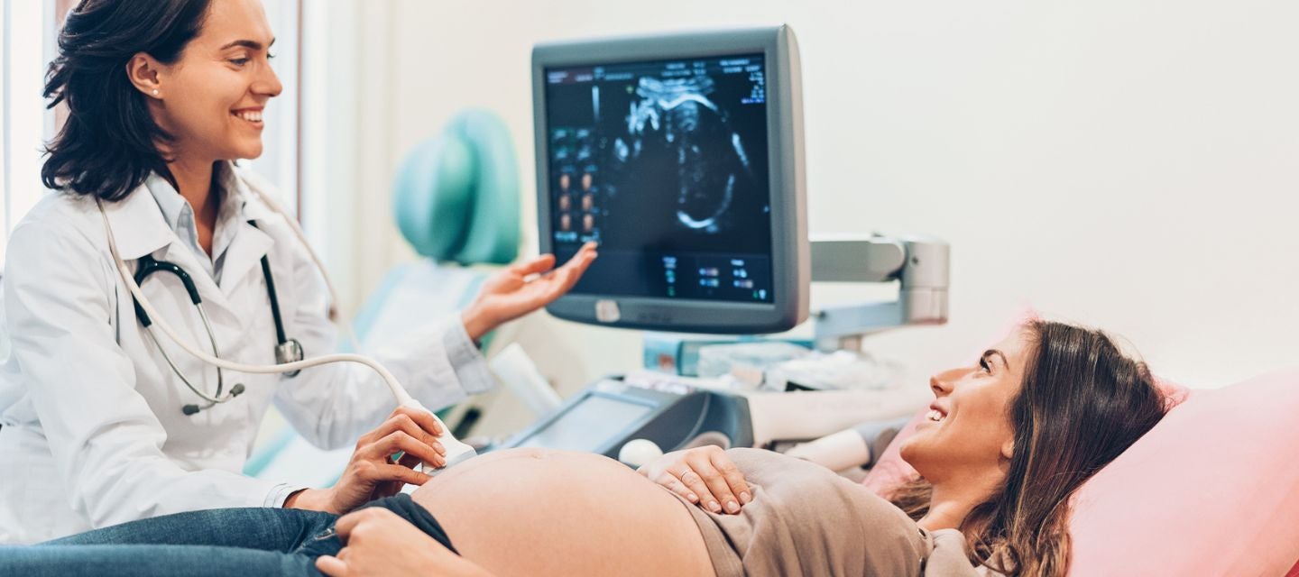
The ultrasound is carried out in real time, so the images you see on the screen show what is happening inside your uterus at that moment, like watching a movie.
The experience of seeing your unborn baby is exciting and positive, and the sonographer carrying out the examination will generally point out easily recognised parts of the body. You might not recognise or understand some of the images you see on the ultrasound screen, but it is all part of this important and thorough screening.
At the 18-22 week scan
Many people wish to know the sex of the foetus, as it can usually be seen at this time. If you would like to know, ask the sonographer to tell you. Occasionally, the sonographer will not be able to tell, usually because of the position of the foetus. If it is not possible to tell the sex, you will not receive another screening ultrasound for that purpose. You should be aware that assessment of the sex is not 100% accurate. If you do not want to know the sex of the foetus, tell the sonographer before starting the ultrasound scan.
The ultrasound is carried out for medical reasons to fully check and assess the development of the foetus from head to toe.
A number of measurements of the foetus will be taken (head size, abdomen and bones) to assess the exact size and age of the baby. The screening ultrasound will look at the position of the placenta and whether it is away from the cervix so that is does not block the birth canal during labour. Measurements and pictures of the cervix are also taken to see if there is a risk of premature labour.
Sometimes the foetus might not be in an ideal position to see a particular structure or part of the body, and the sonographer might ask you to move slightly by rolling from one side to the other. Occasionally, the foetus is in such a position that an area cannot be seen, and you might be asked to return on another day to complete the screening. This should not alarm you and often happens.
The person carrying out the screening ultrasound will be concentrating very closely on the images as they come onto the screen and might be quiet or not talking. Do not be concerned, as they are concentrating on this complex examination.
Pregnancy ultrasound is complex, because there are many structures in the developing foetus that need to be checked and measured. A normal foetus will be moving quite a bit during the scan, and it might take a few minutes to get exactly the right image of a hand, foot, the brain, or various parts of the chest or abdomen.
Is the ultrasound safe? keyboard_arrow_down
Diagnostic ultrasound has been used for over 80 years in pregnancy. Ultrasound is considered safe, and there is no known side effects.
What happens after the scan? keyboard_arrow_down
We will send a report of your scan to the person who referred you for your ultrasound (LMC).
if you attended a Hamilton Radiology site, your images will be available to you with the Hamilton My Images app. Please ensure our office staff has your mobile number to access these images. If you do not wish to receive these, please let us know.
How do I access Hamilton MyImages to see my ultrasound images? keyboard_arrow_down
Hamilton MyImages offers secure sharing of Pregnancy Ultrasound scans with family or friends.
- Personal access from any computer or smartphone
- No more carrying CD's or DVD's
Download additional information on how it works here.
Related procedures

This information has been reviewed & approved by Dr Ronald Shnier (I-MED Chief Medical Officer).
Related procedures
This information has been reviewed & approved by Dr Ronald Shnier (I-MED Chief Medical Officer).
How much will my examination cost?
Fees for radiology tests can vary and depend on a number of factors. Please make an enquiry with us by phone or email to get a quote for the service you require. ACC co-payments may apply.
We accept all radiology referral forms.
Waikato
Phone: 0800 426 723
Email: Booking.Waikato@i-med.co.nz
Rotorua
Phone: 0800 466 5642
Email: Booking.Rotorua@i-med.co.nz
Bay of Plenty
Phone: 07 544 5993
Email: Booking.bop@i-med.co.nz
Taranaki
Phone: 06 759 4317
Email: Booking.Taranaki@i-med.co.nz