

Alzheimer's Disease Imaging & Anti-Amyloid Therapy Guide
Alzheimer's Disease Imaging & Anti-Amyloid Therapy Guide
Radiology Pathways
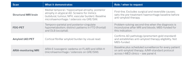
Imaging anchors both the diagnosis of Alzheimer’s disease (AD) and the safe delivery of anti-amyloid therapy. There are four key studies available, which are used in sequence according to the clinical question:
- Structural brain MRI: Recommended for any patient with progressive cognitive decline. It helps exclude structural or reversible mimics and characterises the atrophy pattern (typically medial temporal/parietal/hippocampal in AD).
- FDG-PET: Utilised when the diagnosis remains unclear after an MRI and blood tests. It reads the metabolic signature to separate AD from frontotemporal dementia (FTD) and dementia with Lewy bodies (DLB).
- Amyloid (Aβ) PET: Used to confirm cortical amyloid (the premortem gold standard) and establish patient eligibility for anti-amyloid therapy.
- ARIA-monitoring MRI: Once therapy commences, a baseline scan is required, followed by a scheduled series of surveillance scans to detect amyloid-related imaging abnormalities (ARIA).
Note: While a validated blood test (plasma p-tau217) is emerging as a non-invasive triage step ahead of PET, imaging remains the confirmatory and monitoring backbone.
A Quick Reference Guide
A Quick Reference Guide
Anti-Amyloid Therapy & ARIA
Two amyloid-targeting monoclonal antibodies are now TGA-registered for early symptomatic AD (mild cognitive impairment or mild dementia) with biomarker-confirmed amyloid: donanemab (Kisunla) and lecanemab (Leqembi). Please note: Neither is currently PBS-subsidised.
Eligibility Requirements
- Confirmed amyloid (via Aβ-PET or CSF).
- For donanemab, a low/medium-high tau stratum is required.
- ApoE ε4 testing is required prior to initiation. These agents are not indicated in ε4 homozygotes (who represent roughly 15% of AD patients and carry a markedly higher ARIA risk).
ARIA Risks
ARIA (Amyloid-Related Imaging Abnormalities) is the principal treatment risk associated with these therapies:
- ARIA-E: Vasogenic oedema.
- ARIA-H: Microhaemorrhage/superficial siderosis.
Trial rates for these abnormalities:
- Lecanemab: ARIA-E 12.6%, ARIA-H 17.3%
- Donanemab: ARIA-E 24%, ARIA-H 31.4%
ARIA is usually asymptomatic and detected on routine MRIs, most often early in the treatment course (within the first 6 months), though it can occasionally be serious or fatal.
Clinical Warning: ARIA-E can mimic acute stroke. Carefully weigh the use of thrombolysis and maintain heightened vigilance when reading a head CT for any patient on anti-amyloid therapy.
ARIA-Monitoring MRI Protocols & Scheduling
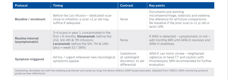
The safe delivery of anti-amyloid therapy depends on standardised serial MRIs. I-MED follows the ASNR/AJNR consensus recommendations (as no separate RANZCR standard currently exists). Every I-MED MRI clinic is trained and configured to deliver the minimum ARIA sequences consistently across our network.
Referral Requirements
Referrals must explicitly state that the patient is on anti-amyloid AD treatment. This ensures the correct protocol (baseline, asymptomatic interval, or symptomatic) and reporting are selected. Non-specific or unclear referrals will be clarified with the treating practitioner prior to scanning.
Minimum Sequences
Our protocols target subtle oedema (FLAIR) and microhaemorrhage/siderosis (GRE/SWI, with DWI), and are kept identical from baseline through to follow-up. Scans are non-contrast for baseline and routine monitoring. Gadolinium is added only at the reporting radiologist's discretion for symptomatic patients.
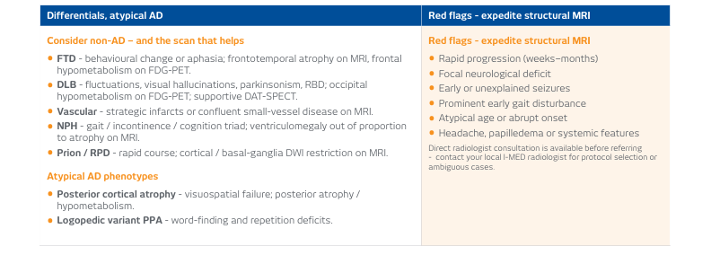
Differentials, Atypical AD & Red Flags
Differentials, Atypical AD & Red Flags
Imaging Access & Medicare Funding (Australia)
- Structural MRI brain: Medicare-eligible on specialist request under the Diagnostic Imaging Services Table.
- FDG-PET: MBS-funded where AD remains inconclusive after comprehensive clinical assessment, MRI, and bloods.
- Amyloid PET: Not currently MBS-funded. Access is via self-funding, clinical trials, or research programs.
- ARIA-monitoring MRI: Repeat monitoring MRIs are not currently Medicare-funded for routine AD management. This must be factored into treatment planning and patient counselling.
Note: Item numbers and funding are subject to MBS review. Please verify currency at mbsonline.gov.au before claiming.